An extracellular network of Arabidopsis leucine-rich repeat receptor kinases

Smakowska-Luzan E, Mott GA, Parys K, Stegmann M, Howton TC, Layeghifard M, Neuhold J, Lehner A, Kong J, Grünwald K, Weinberger N, Satbhai SB, Mayer D, Busch W, Madalinski M, Stolt-Bergner P, Provart NJ, Mukhtar MS, Zipfel C, Desveaux D, Guttman DS, Belkhadir Y

Nature 2018 01;553(7688):342-346

PMID: 29320478

Abstract

The cells of multicellular organisms receive extracellular signals using surface receptors. The extracellular domains (ECDs) of cell surface receptors function as interaction platforms, and as regulatory modules of receptor activation. Understanding how interactions between ECDs produce signal-competent receptor complexes is challenging because of their low biochemical tractability. In plants, the discovery of ECD interactions is complicated by the massive expansion of receptor families, which creates tremendous potential for changeover in receptor interactions. The largest of these families in Arabidopsis thaliana consists of 225 evolutionarily related leucine-rich repeat receptor kinases (LRR-RKs), which function in the sensing of microorganisms, cell expansion, stomata development and stem-cell maintenance. Although the principles that govern LRR-RK signalling activation are emerging, the systems-level organization of this family of proteins is unknown. Here, to address this, we investigated 40,000 potential ECD interactions using a sensitized high-throughput interaction assay, and produced an LRR-based cell surface interaction network (CSI) that consists of 567 interactions. To demonstrate the power of CSI for detecting biologically relevant interactions, we predicted and validated the functions of uncharacterized LRR-RKs in plant growth and immunity. In addition, we show that CSI operates as a unified regulatory network in which the LRR-RKs most crucial for its overall structure are required to prevent the aberrant signalling of receptors that are several network-steps away. Thus, plants have evolved LRR-RK networks to process extracellular signals into carefully balanced responses.

Triphosphate Tunnel Metalloenzyme Function in Senescence Highlights a Biological Diversification of This Protein Superfamily

Ung H, Karia P, Ebine K, Ueda T, Yoshioka K, Moeder W

Plant Physiol. 2017 Sep;175(1):473-485

PMID: 28733390

Abstract

The triphosphate tunnel metalloenzyme (TTM) superfamily comprises a group of enzymes that hydrolyze organophosphate substrates. They exist in all domains of life, yet the biological role of most family members is unclear. Arabidopsis (Arabidopsis thaliana) encodes three TTM genes. We have previously reported that AtTTM2 displays pyrophosphatase activity and is involved in pathogen resistance. Here, we report the biochemical activity and biological function of AtTTM1 and diversification of the biological roles between AtTTM1 and 2 Biochemical analyses revealed that AtTTM1 displays pyrophosphatase activity similar to AtTTM2, making them the only TTMs characterized so far to act on a diphosphate substrate. However, knockout mutant analysis showed that AtTTM1 is not involved in pathogen resistance but rather in leaf senescence. AtTTM1 is transcriptionally up-regulated during leaf senescence, and knockout mutants of AtTTM1 exhibit delayed dark-induced and natural senescence. The double mutant of AtTTM1 and AtTTM2 did not show synergistic effects, further indicating the diversification of their biological function. However, promoter swap analyses revealed that they functionally can complement each other, and confocal microscopy revealed that both proteins are tail-anchored proteins that localize to the mitochondrial outer membrane. Additionally, transient overexpression of either gene in Nicotiana benthamiana induced senescence-like cell death upon dark treatment. Taken together, we show that two TTMs display the same biochemical properties but distinct biological functions that are governed by their transcriptional regulation. Moreover, this work reveals a possible connection of immunity-related programmed cell death and senescence through novel mitochondrial tail-anchored proteins.

A high-sensitivity, microtiter-based plate assay for plant MAMP-triggered immunity

Mott GA, Desveaux D, Guttman DS

Mol. Plant Microbe Interact. 2017 Dec;

PMID: 29199888

Abstract

The first step in the plant immune response to pathogen challenge involves the perception of conserved epitopes, called microbe-associated molecular patterns (MAMPs), by cell surface pattern recognition receptors (PRRs). Given the key roles that MAMPs and PRRs play in plant innate immunity, great effort has been expended to identify these molecules. Current methods for assaying these immune responses are often limited in their resolution and throughput, and consequently, there is a need for medium- to high-throughput methodologies. Here, we describe the development of a 96-well microtiter plate-based assay for plant PTI that measures the activity of plant peroxidase (POX) enzymes produced in response to treatment with bacterial MAMPs. The system has been optimized to minimize both the amount of plant tissue and MAMP required, and displays up to three orders of magnitude greater sensitivity than the traditional luminol-based reactive oxygen species (ROS) assay when measuring the plant response to treatment with the bacterial MAMP flg22, reaching detection limits in the picomolar range. This high sensitivity opens up the possibility of evaluating the immune-eliciting effects of weaker elicitors. The throughput and material requirements of the assay make it ideal for screens involving quantitative measurement of the plant innate immune response to MAMPs.

Oh, the places they’ll go! A survey of phytopathogen effectors and their host targets

Khan M, Seto D, Subramaniam R, Desveaux D

Plant J. 2017 Nov;

PMID: 29160935

Abstract

Phytopathogens translocate effector proteins into plant cells where they sabotage the host cellular machinery to promote infection. An individual pathogen can translocate numerous distinct effectors during the infection process to target an array of host macromolecules (proteins, metabolites, DNA, etc…) and manipulate them using a variety of enzymatic activities. In this review, we have surveyed the literature for effector targets and curated them to convey the range of functions carried out by phytopathogenic proteins inside host cells. In particular, we have curated the locations of effector targets, as well as their biological and molecular functions and compared these properties across diverse phytopathogens. This analysis validates previous observations about effector functions (eg. immunosuppression), and also highlights some interesting features regarding effector specificity as well as functional diversification of phytopathogen virulence strategies. This article is protected by copyright. All rights reserved.

Identification and analysis of seven effector protein families with different adaptive and evolutionary histories in plant-associated members of the Xanthomonadaceae

Assis RAB, Polloni LC, Patané JSL, Thakur S, Felestrino ÉB, Diaz-Caballero J, Digiampietri LA, Goulart LR, Almeida NF, Nascimento R, Dandekar AM, Zaini PA, Setubal JC, Guttman DS, Moreira LM

Sci Rep. Published Nov 23, 2017.
https://doi.org/10.1038/s41598-017-16325-1

PMID: 29170530

Abstract

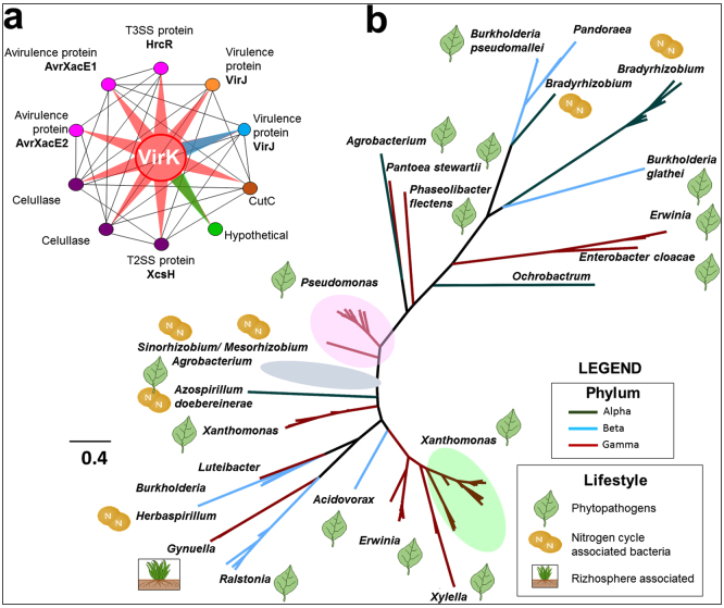
The Xanthomonadaceae family consists of species of non-pathogenic and pathogenic γ-proteobacteria that infect different hosts, including humans and plants. In this study, we performed a comparative analysis using 69 fully sequenced genomes belonging to this family, with a focus on identifying proteins enriched in phytopathogens that could explain the lifestyle and the ability to infect plants. Using a computational approach, we identified seven phytopathogen-enriched protein families putatively secreted by type II secretory system: PheA (CM-sec), LipA/LesA, VirK, and four families involved in N-glycan degradation, NixE, NixF, NixL, and FucA1. In silico and phylogenetic analyses of these protein families revealed they all have orthologs in other phytopathogenic or symbiotic bacteria, and are involved in the modulation and evasion of the immune system. As a proof of concept, we performed a biochemical characterization of LipA from Xac306 and verified that the mutant strain lost most of its lipase and esterase activities and displayed reduced virulence in citrus. Since this study includes closely related organisms with distinct lifestyles and highlights proteins directly related to adaptation inside plant tissues, novel approaches might use these proteins as biotechnological targets for disease control, and contribute to our understanding of the coevolution of plant-associated bacteria.

Convergent selection pressures drive the evolution of rhodopsin kinetics at high altitudes via non-parallel mechanisms

Castiglione GM, Schott RK, Hauser FE, Chang BSW

Evolution. Published Nov 16, 2017.
https://doi.org/10.1111/evo.13396

PMID: 29143302

Abstract

Convergent evolution in response to similar selective pressures is a well-known phenomenon in evolutionary biology. Less well understood is how selection drives convergence in protein function, and the underlying mechanisms by which this can be achieved. Here we investigate functional convergence in the visual system of two distantly related lineages of high-altitude adapted Andean and Himalayan catfishes. Statistical analyses revealed in the two high-altitude lineages, a parallel acceleration of evolutionary rates in rhodopsin, the dim-light visual pigment. However, the elevated rates were found to be accompanied by substitutions at different sites in the protein. Experiments substituting Andean- or Himalayan-specific residues significantly accelerated the kinetic rates of rhodopsin, destabilizing the ligand-bound forms. As found in cold-adapted enzymes, this phenotype likely compensates for a cold-induced decrease in kinetic rates, properties of rhodopsin mediating rod sensitivity and visual performance. Our study suggests that molecular convergence in protein function can be driven by parallel shifts in evolutionary rates but via non-parallel molecular mechanisms. Signatures of natural selection may therefore be a powerful guide for identifying complex instances of functional convergence across a wider range of protein systems. This article is protected by copyright. All rights reserved.

Insights into visual pigment adaptation and diversity from model ecological and evolutionary systems

Hauser FE, Chang BS

Curr Opin Genet Dev. Published Nov 2, 2017.
https://doi.org/10.1016/j.gde.2017.09.005

PMID: 29102895

Abstract

Sensory systems provide valuable insight into the evolution of molecular mechanisms underlying organismal anatomy, physiology, and behaviour. Visual pigments, which mediate the first step in visual transduction, offer a unique window into the relationship between molecular variation and visual performance, and enhance our understanding of how ecology, life history, and physiology may shape genetic variation across a variety of organisms. Here we review recent work investigating vertebrate visual pigments from a number of perspectives. Opsin gene duplication, loss, differential expression, structural variation, and the physiological context in which they operate, have profoundly shaped the visual capabilities of vertebrates adapting to novel environments. We note the importance of conceptual frameworks in investigating visual pigment diversity in vertebrates, highlighting key examples including evolutionary transitions between different photic environments, major shifts in life history evolution and ecology, evolutionary innovations in visual system anatomy and physiology, as well as shifts in visually mediated behaviours and behavioural ecology. We emphasize the utility of studying visual pigment evolution in the context of these different perspectives, and demonstrate how the integrative approaches discussed in this review contribute to a better understanding of the underlying molecular processes mediating adaptation in sensory systems, and the contexts in which they occur.

Staphylococcus aureus interaction with Pseudomonas aeruginosa biofilm enhances tobramycin resistance

Beaudoin T, Yau YCW, Stapleton PJ, Gong Y, Wang PW, Guttman DS, Waters V

NPJ Biofilms Microbiomes. Published Oct 19, 2017.
https://doi.org/DOI

PMID: 29062489

Abstract

Antimicrobial resistance is a significant threat to the treatment of infectious disease. Multiple mechanisms of resistance to different classes of antibiotics have been identified and well-studied. However, these mechanisms are studied with bacteria in isolation, whereas often, infections have a polymicrobial basis. Using a biofilm slide chamber model, we visualized the formation and development of clinical Pseudomonas aeruginosa biofilms in the presence of secreted Staphylococcus aureus exoproducts, two bacteria that commonly co-infect pediatric patients with cystic fibrosis. We showed that, over time, certain isolates of P. aeruginosa can form different biofilm architecture in the presence of S. aureus exoproducts. We further determined that this interaction was dependent on Psl produced by P. aeruginosa and staphylococcal protein A from S. aureus. Importantly, we identified a mechanism of antibiotic resistance to tobramycin that is dependent on the polymicrobial interactions between these two bacteria. This interaction occurred in isolates of P. aeruginosa recovered from children with cystic fibrosis who failed to clear P. aeruginosa following inhaled tobramycin treatment.

Exocyst, exosomes, and autophagy in the regulation of Brassicaceae pollen-stigma interactions

Goring DR

J Exp Bot. Published Sep 27, 2017.
https://doi.org/10.1093/jxb/erx340

PMID: 29036428

Abstract

Brassicaceae pollen-stigma interactions have been extensively studied in Brassica and Arabidopsis species to identify cellular events triggered in the stigmatic papillae by pollen contact. Compatible pollinations are linked to the activation of basal cellular responses in the stigmatic papillae, which include calcium gradients, actin networks, and polarized secretion. The occurrence of these cellular events in stigmatic papillae coincides with the stages of pollen hydration and pollen tube entry into the stigmatic papillar cell wall. However, the form of the vesicle trafficking appears to differ between species, with vesicle-like structures detected in Arabidopsis species while exosomes were found to be secreted in Brassica species. Around the same timeframe, self-incompatible pollen recognition leads altered cellular responses in the stigmatic papillae to interfere with basal compatible pollen responses and disrupt regulated secretion, causing self-pollen rejection. Here, the literature on the changing cellular dynamics in the stigmatic papillae following pollination is reviewed and discussed in the context of other well-characterized examples of polarized secretion in plants.

Cesarean Section, Formula Feeding, and Infant Antibiotic Exposure: Separate and Combined Impacts on Gut Microbial Changes in Later Infancy

Yasmin F, Tun HM, Konya TB, Guttman DS, Chari RS, Field CJ, Becker AB, Mandhane PJ, Turvey SE, Subbarao P, Sears MR; CHILD Study Investigators, Scott JA, Dinu I, Kozyrskyj AL

Front Pediatr. Published Sep 26, 2017.
https://doi.org/10.3389/fped.2017.00200

PMID: 29018787

Abstract

Established during infancy, our complex gut microbial community is shaped by medical interventions and societal preferences, such as cesarean section, formula feeding, and antibiotic use. We undertook this study to apply the significance analysis of microarrays (SAM) method to quantify changes in gut microbial composition during later infancy following the most common birth and postnatal exposures affecting infant gut microbial composition. Gut microbiota of 166 full-term infants in the Canadian Healthy Infant Longitudinal Development birth cohort were profiled using 16S high-throughput gene sequencing. Infants were placed into groups according to mutually exclusive combinations of birth mode (vaginal/cesarean birth), breastfeeding status (yes/no), and antibiotic use (yes/no) by 3 months of age. Based on repeated permutations of data and adjustment for the false discovery rate, the SAM statistic identified statistically significant changes in gut microbial abundance between 3 months and 1 year of age within each infant group. We observed well-known patterns of microbial phyla succession in later infancy (declining Proteobacteria; increasing Firmicutes and Bacteroidetes) following vaginal birth, breastfeeding, and no antibiotic exposure. Genus Lactobacillus, Roseburia, and Faecalibacterium species appeared in the top 10 increases to microbial abundance in these infants. Deviations from this pattern were evident among infants with other perinatal co-exposures; notably, the largest number of microbial species with unchanged abundance was seen in gut microbiota following early cessation of breastfeeding in infants. With and without antibiotic exposure, the absence of a breast milk diet by 3 months of age following vaginal birth yielded a higher proportion of unchanged abundance of Bacteroidaceae and Enterobacteriaceae in later infancy, and a higher ratio of unchanged Enterobacteriaceae to Alcaligenaceae microbiota. Gut microbiota of infants born vaginally and exclusively formula fed became less enriched with family Veillonellaceae and Clostridiaceae, showed unchanging levels of Ruminococcaceae, and exhibited a greater decline in the Rikenellaceae/Bacteroidaceae ratio compared to their breastfed, vaginally delivered counterparts. These changes were also evident in cesarean-delivered infants to a lesser extent. The clinical relevance of these trajectories of microbial change is that they culminate in taxon-specific abundances in the gut microbiota of later infancy, which we and others have observed to be associated with food sensitization.